Friday, February 18, 2011

Ascaris lumbricoides: Biggest you've ever seen

http://diseasepicture.com/ascariasis?pid=94

Introduction: Ascaris lumbricoides is the largest intestinal worm to infect humans. These worms can reach up to 18 inches in length, and survive by ingesting intestinal contents and fluids. These worms are found in areas with poor sanitation and malnutrition. People that have Ascaris lumbricoides have severe gastrointestinal pain caused ascarisis. Symptoms of ascarisis include fever, jaundice, wheezing, and abdominal tenderness due to the movement of worms through intestines. This disease is not a deadly disease but has caused 8,000-100,000 deaths due to gastrointestinal complications, such as ruptures and blockages. [1]

Symbiont Description: Ascaris lumbricoides belongs to the phylum Nematoda, and has a smooth hairless body. The males are relatively smaller than the females. Males are normally 7.8 in while the females are 12 in [2]. The males also differ from the females because their tails have a hook shape and tethered ends. The mouths of A. lumbricoides have three lips with very sharp teeth that assist in attaching to the intestinal wall and movement throughout the body [2].


http://www.nematode.net/NN3_frontpage.cgi?navbar_selection=speciestable&subnav_selection=Ascaris_lumbricoides

Host Description: A. lumbricoides only host is humans, but mostly children. Children have a higher risk of getting infected because they are more likely to play in soil than adults. Also boys are more likely to get infected than girls. When comparing US to the rest of the world, 4 million Americans have been reported to have these worms. On an international level 1.4 billion people have been infected with these worms [1]. Some patients have been known to have only a few worms which are easily treatable. But in some cases there can be hundreds of worms in patients causing extreme pain and gastrointestinal trauma [3].

Life Cycle:


The process starts by coming into contact with A. lumbricoides infective eggs, by ingesting soil or crops that have not been washed properly. Once inside the body the eggs hatch and release larvae to penetrate the intestinal wall. Larvae migrate to the pulmonary system and then to the alveolar area causing the host to cough. After 1-2 weeks a cough allows the larvae to be swallowed and sent in the direction of the small intestines. These larvae mature into adults and lay eggs. The eggs are excreted through the feces. Once the eggs are excreted it takes about 1 week for them to become infective eggs and is able to infect another host. The full process from egg to adult takes 9 weeks [7]. Adult worms can live 6-24 months in a host. Since these worms live in the human body for up to 2 yrs they have been known to migrate to different areas of the body, mostly the nose, mouth, and anus [1]. Some patients have reported waking up with a pillow full of worms. What a way to wake up!!!.

Ecology: A. lumbricoides are found in tropical and subtropical areas because of the humid and warm conditions that aid in the survival of the eggs. These worms have also been reported in areas where human waste is used as fertilizer, and in the southeast areas of the US. Most infections happen in Latin America (8%), Africa (12%), and Asia (73%) [3]. Treatment of this disease for acute cases is through albendazole, ivermectin, or mebendazole. These medications work by killing the adult worms. In major cases, more than 20, surgery is needed to excrete the worms from the body and repair the damage they have caused [4] [5].

An example of Ascaris lumbricoides: This is an example of how a parasite has a complex life cycle to survive in the human body. The larvae have to develop in a secure area, alveolar area, so that they can grow to withstand the acid of the stomach. Since this process takes up to 9 days this is important. If the larvae try to leave to quickly they can be destroyed by the stomach acid. The complex life cycle also increases fitness because the females are able to reproduce and lay eggs that are able to infect another host, which in turn keeps the cycle going.

References :
[1]- http://emedicine.medscape.com/article/788398-overview
[2]- http://chestofbooks.com/health/disease/Intestines/Ascaris-Lumbricoides-Common-Spool-Or-Round-Worm.html
[3]- http://www.humanillnesses.com/original/A-As/Ascariasis.html#ixzz1EElwuXri
[4]- http://www.mayoclinic.com/health/ascariasis/DS00688/DSECTION=treatments-and-drugs
[5]- http://www.metapathogen.com/roundworm/
[6]- http://www.suite101.com/content/ascaris-lumbricoides-life-cycle-a32510
[7]- http://www.dpd.cdc.gov/dpdx/html/ascariasis.htm

Trichinella Spiralis - Trix are for pigs!


Trichinella Spiralis:
Trichinella species are the smallest nematode parasites of humans. Trichinella Spiralis, more commonly known as a threadworm or pork worm, is a nematode responsible for the disease trichinosis or trichinellosis [2]. It is usually caused by the consumption of unsanitary, undercooked , and infected meat, especially pork. Trichinella lodges itself in striated muscle cells. An estimated 10,000 cases of Trichinosis occur every year worldwide [5]. The disease is most commonly reported in animals rather than humans.

Symbiont Description:
Trichinella spiralis is part of the family Trichinellidae in the order Trichurida. Trichinella males measure between 1.4mm and 1.6mm long with a terminal anus [1]. Females of these species are two times as big as the males, also with a terminal anus. The esophagus of the female contains the vulva and the uterus in the posterior end contains developing eggs [1]. The anterior end of the nematode contains developed juveniles.


Host Description:
Species of Trichinella spiralis are most commonly found in pigs, rats or other wild game such as bears or cougars. Domestic pigs are very effective hosts and rats are usually the intermediate hosts. Humans, however, are considered accidental hosts. The reason for this is because humans cannot carry the parasite on to the next life cycle [6]. Pigs and rats, on the other hand, are able to carry the parasite on to its next life cycle by eating infected meat.


Life Cycle:
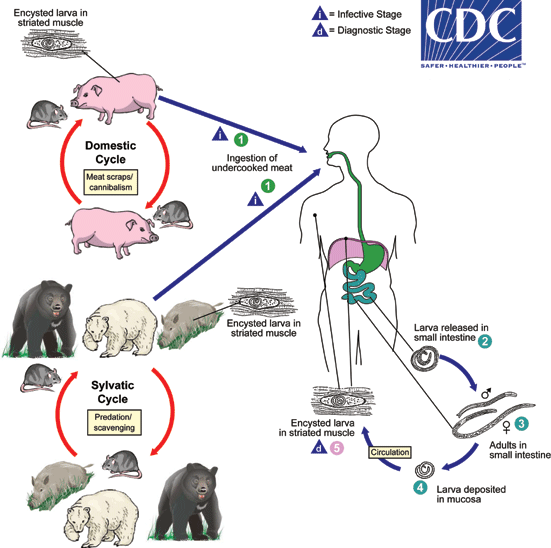
Trichinosis is caused by consuming infected meat containing cysts of Trichinella spiralis. These cysts are also known as nurse cells. As the cyst containing meat travels to the stomach, chemical signals caused by pepsin allow the cyst to release the larvae [6]. The meat then enters the intestine, where the chemical agent bile allows the larvae to move in a snakelike slither manner [6]. The larvae leave the meat and enter the intestine where they become adults. After one week, the adult females release larvae that find its way through the blood and lymphatic system to striated muscle cells where they lodge and form cysts. The female then dies and passes out of the host [1].The cycle is repeated once again when muscle meat containing cysts is consumed.

Ecology:
This parasite can be harmful to the host. In humans, Trichinella can cause minor symptoms such as nausea, vomiting, diarrhea, and abdominal pain [5]. Many of the symptoms of Trichinella are assumed to be a common illness such as the flu [5]. The parasite is most commonly diagnosed by a blood test and rarely through a muscle biopsy [5]. Cases of this parasite have decreased due to freezing meat. When meat such as pork is frozen, the larvae inside the cysts are killed.

An Example Of:
Trichinella uses a variety of chemical triggers to further its life cycle. The parasite is highly complex and uses chemical agents found inside the body to encounter the host. Two specific agents the parasite uses to release larvae into the host are pepsin and bile [6]. These chemical agents help the fitness of the parasite by allowing it to complete and repeat its lifecycle.

References:
1. http://en.wikipedia.org/wiki/Trichinella_spiralis
2. http://www.e-cleansing.com/parasites/threadworm-trichinella-spiralis-2.html
3. http://www.trichinella.org/
4. http://www.wormbook.org/chapters/www_genomesTrichinella/genomesTrichinella.html
5. http://www.cdc.gov/parasites/trichinellosis/
6. Zimmer, Carl. Parasite Rex. London: Arrow, 2000. Print.

Wednesday, February 16, 2011

Giardia lamblia– The Intestinal Parasite


Giardia lamblia is a single-celled protozoon. This parasite moves with the aid of five flagella. Giardia lamblia causes Giardiasis which is the most frequent cause of non-bacterial diarrhea in North America. This is the most common intestinal parasite disease in North America. Many developing countries come into contact with this parasitic disease. Some symptoms that one may experience include diarrhea, abdominal pain, nausea, gas, and fever. For some children, this disease can affect their physical and mental growth, slower their development, and cause malnutrition. [1]


Giardia lamblia
Giardia lamblia is a rename for Giardia intestinali and Giardia duodenalis. It was renamed in 1915 in honor of Professor A. Giard of Paris and Dr. F. Lambli of Prague. [2]. The parasite is from the phylum Metamonada which is the group of flagellate protozoa. The genus Giardia is widespread and infects many different species. [5]
The genome of the parasite consists of five chromosomes and the sizes range from 0.7 to over 3 Mb. It has a tear-drop shape and measure ten-fifteen um in length. Their shape is important for differentiating between species. These parasites do not consist of mitochondria, endoplasmic reticulum, or lysosomes. [2]

Host Description
This parasite is widespread because it infects humans and other mammals. Many children are infected by this parasite also. It infects about 6-8% of children and 3 % of adults. People come into contact with parasite, most often, through contaminated water. This disease may be passed on when a human contacts another human or an animal. Men who have sex with other men are at a greater risk of getting infected. Just like other diseases, travelers are most likely to be at the greatest risk because of the traveling to other countries where Giardia might be common. [1]

Life Cycle:
The Giardia lamblia consists of a two-stage life cycle. It contains the trophozoite and cyst. Cysts can survive in water for ever several months.
• Contaminated water, food, or hands with cysts is ingested by people of mammals.
• In the small intestine, excystation occurs and releases trophozoites. One cyst produces two trophozoites.
• Trophozoites multiply by binary fission in the small bowel where they can be free or attached to the mucosa by ventral sucking disks.
• In the colon, encystation occur and cysts become infectious.
Cysts are the most common form found in the nondiarrheal feces. Since the cysts are infectious in the stool, person-to-person transmission is possible. [1]. The main food source for this parasite is glucose which is obtained by diffusion or pinocytosis. [2]


Ecology
Giardia is worldwide. It can be found in soil, water, food, and surfaces that have been contaminated with feces from infected humans or animals. [2] The parasite cannot be transmitted and infect someone through the contact of blood. They don’t invade tissues but adhere closely to the lining of the small intestine [2]. Cool, moist areas increase the survival of these organisms [3]. Statistics have also shown that infection rates are higher in the summer than any other time of the year.
There is treatment available for this parasitic disease. Some medications that can help treat the disease include metronidazole, tinidazole, and nitazoxanide. [1]


An example of Giardia lamblia
Giardia lamblia can be an example of how the host has both lines of defense: for encounter and compatibility. The host has defenses that will help it from encountering the parasites. One can close the encounter filter through better hygiene and education. If people do not drink contaminated water or eat contaminated food or clean surfaces, then the spread of the parasite would lessen. This will help avoid the parasite from contacting humans and other mammals. If the first line of defense works, then the parasite is destroyed in the environment and the host does not get infected. However, if the host does encounter the parasite, then it needs to find a compatibility filter. Here the parasite can be fought through vaccination and treatment. There are several medications that help treat Giardia lamblia. These defenses decrease the parasite from spreading worldwide. [4]


Websites
[1] - http://www.cdc.gov/parasites/giardia/gen_info/index.html
[2] - http://microbewiki.kenyon.edu/index.php/Giardia
[3] - http://www.fda.gov/food/foodsafety/foodborneillness/foodborneillnessfoodbornepathogensnaturaltoxins/badbugbook/ucm070716.htm
[4] – Combes, Claude. The Art of Being a Parasite. Chicago: The University of Chicago, 2005. 12-15.
[5] - http://en.wikipedia.org/wiki/Giardia#Systematics

Tuesday, February 15, 2011

Strongylus vulgaris: A Deadly Traveler

Also known as a bloodworm or redworm, Strongylus vulgaris is a dangerous parasitic roundworm of horses [1]. It has a global distribution, found in grasslands of all temperate regions of the world. This organism migrates throughout the horses’ arterial system for at least 4 months before it returns to the intestines to mature and reproduce [2]. While travelling, the nematode larvae damages mesentery and arterial walls, leading to various complications, usually boot clots [1], and diseases such as Adams’ Lameness (spinal nematodiasis) [4]. This parasite is found in areas where the horse population is heavily concentrated [1], and before effective medications were developed, it could be found in 90 to 100 percent of horse in the United States [3].


Symbiont Description

Strongylus vulgaris is a small parasite with the adult males ranging from 2.5 to 4 cm and the females ranging from 4 to 6 cm [3]. This stout worm is dark red in color due to the ingestion of blood from the host [5]. These worms do not have extensive social behaviors, with most of the interactions occurring in the intestines when mating is occurring [3]. It uses a buccal capsule with two hooked teeth to attach and feed on the intestinal wall, looking very such like hookworms found in humans. Its cuticle is made up of three or more layers secreted by the epidermis that aid in the invasion of a horse’s digestive tract. Also there are muscles that run longitudinally along its body.


Host Description

Because horses are found I most regions of the world, they remain susceptible to encountering this parasite. Horses also tend to travel in herds or are kept in groups, which increases the likelihood of becoming infected. Horses obtain this parasite from grazing in grass that has been contaminated by larvae, either from feces or even the soil. As long as there is not a period of drought and horse droppings are not broken up, the bloodworm will continue to be present in any given grazing field [1].


Life cycle

The reproduction begins when the female bloodworm releases pheromones to attract males [3]. The male then curves around the female genital opening and uses spicules to hold the female while his sperm swim to fertilize the females’ eggs [3]. Then the life cycle begins [1]…
 Eggs that are produced are passed with the feces of the horse. They remain in these moist and warm conditions where they soon hatch. It takes about two weeks for the larvae to reach the infective stage.
 When ingested, the larvae penetrate the lining of the intestines.
 They eventually migrate to the arteries where they burrow along the walls. This stage can occur for up to three months.
 During this time, they become “pre-adults”, ready to travel back to the intestines, where they form nodules in the intestinal arteries.
 After feeding for about 8 weeks, they reach sexual maturity and begin producing eggs, all the while sucking blood from the horse.

Ecology

The ecological impact of these organisms has the most influence on any horse population. It can cause death if untreated, which is usually the case for populations of wild horses [5]. It has an economic impact on horse farms. Many horse breeders closely watch the area that their horses graze. Once the parasitic larvae penetrate the intestinal wall, it migrates through the mesentery arteries. As it travels along the walls, S. vulgaris causes damage, most significantly lesions. These lesions can become severe enough to thicken the arterial walls resulting in blood clotting and eventually thrombosis. Furthermore, parts of the clots can break off, travel, and clot other vessels throughout the body. Death occurs when impairment of the blood supply in the intestines causing colic and the development of gangrene.


A Rendezvous in Space and Time

When in a contaminated field, S. vulgaris will continuously be present because it remains in its prime spot. It takes advantage of the free living stage of its life cycle to crawl on to the grass blades when the horses will be out to graze. There is no intermediary meaning no chance to get lost on its way to the final post. On the other hand, because it employs a 6 part life cycle, it increases the fitness of the organism. It spends months traveling around the body developing, never being in the same spot for a long period of time. Then it uses it morphological features (hooked head) to latch on to feed and reproduce for and even longer period of time.


References

[1] http://www.suite101.com/content/strongylus-vulgaris-bloodworm-a33705
[2] http://cal.vet.upenn.edu/projects/merial/Strongls/strong_8d.htm
[3] http://animaldiversity.ummz.umich.edu/site/accounts/information/Strongylus_vulgaris.html
[4]http://books.google.com/books?id=MQTRQvwY2MQC&pg=PA1074&lpg=PA1074&dq=spinal+nematodiasis&source=bl&ots=IGQthyt_-3&sig=Ui2O7rq2pIJGyDuGFXA_lhNRzxI&hl=en&ei=Nh1bTaPmK4bGlQfygdHkDA&sa=X&oi=book_result&ct=result&resnum=1&ved=0CBcQ6AEwAA#
[5] http://ph.merial.com/equine/pdf/equine_vteq_lgst.pdf

Monday, February 14, 2011

Dirofilaria immitis: Eat your heart out


Dirofilaria immitis, although it is just another parasitic organism, grabs our attention more than any other parasite, especially those of you who have pets. We see signs in pet stores and watch commercials on T.V. that tell us the dangers of dog heartworm. Transmitted by mosquito bites, heartworm, Dirofilaria immitis, is a common filarial parasitic nematode that infects primarily dogs and other canine species. It usually resides in the arteries, heart, lungs, as well as other blood vessels in dogs. Heartworms cause serious vascular damages and can be fatal, especially in working dogs with high levels of physical activity. Heartworm can be found all over, prevalent in areas with mosquitoes[1].

SYMBIONT DESCRIPTION:
Heartworms belong to class of nematoda called secernentea. Dirofilaria immitis is characterized by its cylindrical threadlike body with a tough exterior. A sharp spear at the top of their heads is used to puncture cells. Both male and female heartworms have similar characteristics but differ in size. The female worm is 10 to 12 inches long while males are half the size of the female. They receive their nutrients from the red blood cells surrounding the heart and lungs of their hosts. An adult female worm can release up to 5,000 larvae per day into the blood stream[2]. One interesting fact about this parasite is that it carries endosymbiotic Wolbachia bacteria, whose metabolites affect the host's immune response[3].

HOST DESCRIPTION:
Dirofilaria immitis is capable of infecting a wide variety of mammals. While dogs are considered the definitive host of heartworms, they can infect more than 30 species including cats and humans (very rare). Most infected dogs do not show any signs of disease for as long as two years. Unfortunately, by that time the disease is well advanced. The signs of the disease depend on the number of adults present and the degree of damage done to the heart and other organs. Few of the most obvious signs are: chronic cough, shortness in breath, loss of stamina and weakness. All of these signs are most noticeable following exercise, when some dogs even faint from lack of air supply to their lungs. The disease is diagnosed mostly in four to eight year old dogs. Moreover, various mosquito species are intermediate hosts of Dirofilaria immitis larvae. They are capable of supporting larval development to the infected larval stage as well as transmitting the disease[4].

LIFE CYCLE:
Knowing the life cycle and how Dirofiliaria immitis reproduces is a key factor in understanding why this parasite is such a problem. The life cycle begins when a female black mosquito takes a blood meal from an infected host. The blood contains microfilariae, long thin embryos of Dirofilaria immitis, that look like tiny worms. Microfilariae lives about two weeks in the mosquito salivary gland before becoming inactive. It develops to the infective larval stage inside the mosquito and migrate within the insect's body to its mouth parts[1]. Next time an infected mosquito bites a dog, cat or any canine, heartworm larvae drops onto the host's skin and migrates in through the wound. In the dermal layer of the skin, and the muscle tissue, the larvae continues to mature for eight to ten weeks, attaining a length of about 25mm as they migrate through tissue heading to the heart and lung vessels[5]. Within 70 to 90 days, the larvae finally reaches the right side of the lungs and the pulmonary artery, where they impede blood flow and cause damage to the heart and lung vessels. Proving both male and female worms are present, Dirofiliaria immitis adult worms mate and females produce eggs, which develop into the long thin microfilariae that are released into the bloodstream. The offspring are produced 7 to 9 months after the mosquito bite. Is it these microfilariae that mosquitos take in when they draw blood from the infected host and the cycle starts all over again. Cats rarely have this microfilariae circulating for the female mosquitoes to pick it up[1].

ECOLOGY:
Heartworms are primarily found in the right ventricle of its host's heart. On a geographical level, Dirofilaria immitis occurs all over the world, including 50 states in America, where there are a large number of mosquitoes. Mosquitoes thrive in warm and wet conditions, therefore tropical countries and regions that are closer to the equator have a greater presence of Dirofilaria immitis. The maturation of larvae ceases when the temperature drops below 57oF but the transmission never reaches zero in winter. Moreover, from an ecological standpoint most of the third world and other developing countries are not capable of medicating host's affected with this parasite because they are not able to afford the medication[1].

AN EXAMPLE OF......
Dirofilaria immitis is an example of a type C parasite that uses vectors or biting organisms to inject themselves through the host integument. Vectors are considered to be invertebrate animals, usually arthropods and in this case mosquitoes are the intermediate species. The pathogen multiplies within the arthropod vector and transmitted when the arthropod takes a blood meal. Furthermore, what is remarkable in these Dirofilaria immits is the usage of double strategy of manipulating both space and time to enhance fitness. Only the microfilariae in the peripheral blood vessels is transmitted when the mosquito takes a blood meal. Also, mosquitoes are nocturnal, only bite in the evening and at night. Therefore, because microfilariae can only be ingested during nighttime, the number of microfilariae present in the peripheral vessels rises at nightfall[6].

REFERENCES:

[1] http://www.heartwormsociety.org/veterinary-resources/canine-guidelines.html
[2] http://www.dailypuppy.com/articles/characteristics-of-heart-worms/2560dfe7-db05-2b83-1c45-559394c117bc
[3] http://www.metapathogen.com/heartworm/
[4] http://docs.google.com/viewer?a=v&q=cache:7tbRlnXIqVwJ:www.vetgateglobal.com/images/usr/191/
canine%2520heartworm%2520disease.pdf+where+are+heartworms+found&hl=en&gl=
us&pid=bl&srcid=ADGEESiArZdqfIvmMQx5l1vuUlFKGpeGz5WJSADkcgoK6mPE-poNZ79-j5x2DavweDqQOYrD6Z7QeruNO5Hl6C_EWCNGcBAjzicmaXWQ_71-l5F_ILlGj5-tGbm1KvgCHQQoMOkw4Vc7&sig=AHIEtbQoTiOA2HimFSZvjUVJS6KhuCic8A
[5]http://www.suite101.com/content/dirofilaria-immitis-heartworm-a21925
[6] Combes, C.(2005). The Art Of Being A parasite. University of Chicago

Sunday, February 13, 2011

Loa loa: A Parasite Worth Seeing


The Loa loa is a nematode, found only in the rainforests and swamps of Central and West Africa [1], and causes causes a disease known as loiasis. The filarial adult worm migrates to the lymphatics [2] and subcutaneous tissues (the third layer of the skin); it may also migrate to the eye under the conjunctiva [3]. Humans become infected when bitten by Chrysops silacea and Chrysops dimidiate (two species of Mango fly) [4].

Description
Adult Males are 20-34 mm long by 350-430 um wide, while females are 20-70 mm long and about 425 um wide [3]. The female vulva is about 2.5 mm from the anterior end, and the tail is about 265-300 um long. Both genders of the L. loa live anywhere from 4 to 17 years [1]. The worm has a simple head, a characteristic of nematodes, with no lips, eight cephalic papillae (sensory organs around the mouth), a long, slender body; and a blunt tail [3].

Host
The definitive host of the worm is humans, whom receive the parasite via its vectors Chrysops silacea and C. dimidiate. Infected mango flies pass on their infected larvae to humans. In turn, a healthy mango fly becomes infected from an infected human [4]. A more detailed description of the process of human infection, and Loa loa life cycle is given below.

Life Cycle


When the Mango Fly bites a human to take a meal Mango Fly larvae are left on the skin and enter through bite.Once inside the body the infective larvae wonder through the subcutaneous tissue while they develop into a mature adults. Adults produce microfilariae measuring 250 to 300 μm by 6 to 8 μm, which are sheathed and have diurnal periodicity (circadian rhythms). Microfilariae have been recovered from spinal fluids, urine, and sputum. During the day they are found in peripheral blood, but during the night, they are found in the lungs.

When another fly ingests microfilariae during a blood meal, the microfilariae lose their sheaths and migrate from the fly's midgut through the hemocoel to the thoracic muscles. It is here that the microfilariae develop into infective larvae. The infective larvae migrate to the fly's proboscis where they can infect another human when the fly takes in a meal.

The Painful Interaction
Worms often go unnoticed as they travel through subcutaneous tissues but can be painful as they pass over the eyeball or bridge of the nose. Swelling of the conjunctiva and eye lid are other characteristics of infection.Patients may describe literaly seeing something crawl across their eye [4]. While in the subcutaneous tissues, the worms trigger immune responses that result in swelling of specific areas often a hand or foot. When they remain in one spot for a short time, localized Calabar swellings, especially in the wrist and ankles, appear, which disappear when the worm moves on. There may also be intense itching, joint pain and fatigue [3]. Other symptoms may include: lymphadenitis (infection of the lymph glands),kidney disease, peripheral neuropathy (damaged peripheral nervous), and retina damage [5]

Class-Like Example
The Loa loa worm is a prime example of a parasite that is increasing its "encounter" filter, enabling it to infect a new host. Emergence rhythms allow for maximizations of encountering a host [6].Because humans are greatly dispersed and always on the move, the worms must develop strategies to make the most of their energy. By only coming to the surface of the skin during the day when the flies are feeding, the worms increase their protection by retreating back within the body where they are less likely to be removed.

Removal
Once the Loa loa worm has made its way into the eye, it can be surgically removed. Want to check it out, or is this parasite really not worth ever seeing? You decide, but the video is at the end of this article:

http://www.examiner.com/infectious-disease-in-national/loa-loa-the-african-eye-worm-video

References
1.http://www.itg.be/itg/distancelearning/lecturenotesvandenendene/41_Filariasisp5.htm
2.http://www.dpd.cdc.gov/dpdx/HTML/Filariasis.htm
3.http://course1.winona.edu/kbates/460prog/Loaloa.htm
4.http://www.stanford.edu/group/parasites/ParaSites2006/Loiasis/Index.html
5.http://www.parasitesinhumans.org/loa-loa-eye-worm.html
6.Combes, C.(2005).The Art of Being A Parasite. University of Chicago.


Wednesday, February 9, 2011

Guignardia bidwellii - Return of the Mummy


[1]


Introduction

While some parasites have a direct influence on human populations because they use humans for hosts, others have an indirect influence. Guignardia bidwellii is one such parasite. This fungus is the cause of the disease known as black rot of grapes, and can have a severe economic impact on vineyards and other elements of the wine-making industry, as it can – if left unchecked – destroy entire fields of grapes. [3]

Symbiont Description

Guignardia bidwellii is a fungus that prefers warm, moist climates. It is native to North America, but has spread to Europe, South America, and Asia. [4] Guignardia bidwelli is a member of the genus Guignardia, all known members of which are plant pathogens. Other members of the genus cause black spot of citrus, Freckle of banana, and similar diseases. Guignardia are ascomycotes, or sac fungi.

Host Description

While other members of the genus infect different plants, Guignardia bidwellii causes disease in grapes. Most grape varieties are susceptible to infection, although the degree of susceptibility varies from cultivar to cultivar. The disease affects the entire plant, though it causes the most prominent damage in the fruits and leaves of the infected grape. [1]

[1]


Life Cycle

As shown in the above image, the life cycle of Guignardia bidwellii is relatively simple in that it uses only one host. Infection begins when the fungus’s ascospores are released into the air, carried by raindrops landing on a previously infected fruit. The ascospores are carried to a nearby uninfected plant. When the ascospores land on a developing fruit cluster, the actively infected berries will eventually become shriveled and black; these are referred to by grape growers as “mummies”. Each mummy contains the fruiting bodies of the fungus, called perithecia and pycnidia. The perithecia will winter in the mummy and develop ascospores that are ready to be released during the rains in the following spring.

Should the ascospore land on a growing leaf or shoot, their developmental course will vary slightly from the process that has been described. There the ascospores will form lesions; within each lesion are clusters of pycnidia. Rather than going dormant for the winter, these pycnidia will produce pycnidiospores, which disperse to infect additional plants. Eventually these pycnidiospores will land on a fruit cluster and the mummy-and-ascospore formation process will begin again. [3]


[3]


Ecology

The most prominent feature of Guignardia bidwellii’s ecological impact is its effect on grape growers. As is described in the life cycle, during its infectious stage the fungus causes clusters of infected grapes to shrivel; they are then useless to the grower. In some regions, such as the Ohio River basin, where there is a high level of humidity, it was formerly impossible to grow grapes commercially due to the prevalence of the fungus. Although chemical controls have been developed that are effective against the fungus, it still poses a significant threat to the crops of organic farmers. [3]

An Example Of: How A More Complex Life Cycle Can Enhance Fitness; Use of Vectors

As described above the “mummies”, or infected grape clusters, shrivel and remain on the grape vine over the winter. Once rainfall begins, the raindrops become the fungus’s vectors, releasing stored spores that infect new regions of the same plant or neighboring plants. [2] These traits combined allow a maximum spread of infection during climate conditions favorable to the fungus and allow it to establish itself.

References

1. http://ipm.illinois.edu/diseases/series700/rpd703/

2. http://dailyparasite.blogspot.com/2010/12/december-31-guignardia-bidwellii.html

3. http://ohioline.osu.edu/hyg-fact/3000/pdf/HYG_3004_08.pdf

4. http://www.nysipm.cornell.edu/factsheets/grapes/diseases/grape_br.pdf