Monday, April 9, 2012

It Takes Two, Baby: Fungus and Algae


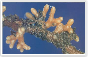
Lichens can easily be found on the side of trees(http://rutgers-leslie.blogspot.com/2010/04/lichen-on-your-trees.html)
Introduction
Lichens are the most well known examples of symbiosis between fungi and plants. Classifications of lichens include some species of mushrooms, slime molds, and some members of the Zygomycota [4]. However, the mutualistic relationship in lichens is basically defined by its association between a fungus and an alga that develops into a unique morphological form that is distinct from either partner [1]. The fungus is called a mycobiont and the algal part is referred to as the phycobiont [1]. The life cycle begins when these components combine, and the fungal filaments enclose to develop into the algal cells. This algal cells provide the lichen with its physical structure and shape. As a result of fungal reproduction, the apothecium produces spores to continue the life cycle again[5]. Like other green plants, it relies on photosynthesis to produce food. The alga contributes food supply through photosynthesis, while fungus protects the alga from losing moisture, harmful solar radiation, and provides the alga with water and nutrients[4]. Although lichens are resilient enough to survive and exploit hard substrata like rocks and tree trunks, they still remain sensitive to air contaminants of the environment.   are so tough that they can survive on hard surfaces such as exposed bare rock surfaces, they are also very sensitive to modern elements introduced to the environment, like sulfur dioxide and other air pollutants [4]. Lichens grow in three forms: foliose or leaf-like, fruticose, that are branched or bushy in appearance, and crustose, which grows a thallus or thin crust upon the surface[2]. Although lichens have the ability to grow in a number of diverse areas, they are most prominent in the Arctic tundra and Antarctic [5]. The global distribution of lichens depends on physical factors like light availability, humidity, substratum nature and chemistry, and sulfur dioxide levels in the air [3]. 
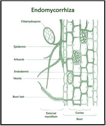
Cross section view of lichen 
                                      (http://www.saburchill.com/ans02/chapters/chap010.html)
Description of the Relationship
 Lichens are dualistic organisms, composed of a fungi and an alga growing together to form single body[2]. The fungal component is often a species of Ascomycetes, although a few of the Basidiomycetes will sometimes grow with green algae such as Protococcus, Cryptococcus, or Trebouxia, or blue green algae such as Gloeocapsa, Nostoc, or Stigonema [5]. The lichen’s method of reproduction can be fungal or asexual[5]. Lichens fungally reproduce by growing fruiting bodies and spores. These spores can produce additional fungi, however, the alga does not get the opportunity to reproduce at all. To continue to life cycle, the new fungus has to either find an algal partner or it perishes [5]. The lichen reproduces asexually by producing soredia, which are small specialized fragments of thallus consisting of fungal tissue. The soredia forms in the parent thallus, grows out through the surface, and are carried by the wind and rain to new environments as small bits of tissue [2]. Lichens are not considered one single organism, but are rather a combination of two organisms that share an intimate relationship[2]. Although fungi and algae that produce lichens can exist in nature alone, many lichens include a fungus that cannot survive with its algae counterpart and takes on a whole new morphology as a separate entity[2]. Based on the anatomy of the lichen, there is an obvious mutualistic symbiosis between the phycobiont and mycobiont [4]. The alga produces the food material and the fungus protects alga from drying out, acquiring too much solar radiation, physically injury, while providing it with water and minerals[4]. Scientists believe the relationship developed because neither component could obtain all the necessary nutrients for survival until they interacted[4].
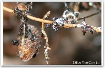
Crutose lichen(http://www.perspective.com/nature/fungi/lichens.html)

Cost/Benefit Analysis
Although fungi and algae only prefer moist and wet environments that do not receive direct sunlight, and cannot survive outside these conditions, Lichens have the ability to grow all over the world [4]. These even include arctic and hot, dry desert areas where not many organisms are able to survive[4]. The alga contributes food supply, and the fungus protects the alga from losing moisture, harmful solar radiation, and provide the alga with water and nutrients [4]. Also, the fungus is able to grow on bare rock and other surfaces where other plants cannot because because the fungus is able to take a firm hold where most plant roots are unable to penetrate[6]. Neither the fungus nor the alga have the ability to solely survive in such hostile environments[6]. However, working as a unit, they can successfully compete with other plants for light and space [6]. Because of the absences of roots, lichens use air as a the primary source of elements[8]. This, however makes them especially vulnerable to industrial air contaminants such as sulfur dioxide[6]. This vulnerability is also correlated with the energy needs of the mycobiont. As the dependency between the mycobiont and phycobiont increases, so does the lichen's vulnerability to air pollution[7,8]. The phycobiont may also use its metabolic energy to repair cellular structures that would otherwise be used towards the maintenance of the photosynthesis process[7,8]. Deviations from equilibration between the mycobiont and phycobiont can lead to complete breakdown of the symbiotic relationship[7]. Therefore the decline in the lichen population may not only be attributed to air contamination, but also from a symbiont that receives more nutrient supplies over the other[8]. The lichen association is in tight knit mutualism in which neither the fungus or algae can reproduce without the balanced efforts of it symbiotic partner.
 
Different faces of the lichen
                                            http://www.youtube.com/watch?v=dHpruP_AaKc

 References
1. http://www.ucmp.berkeley.edu/fungi/lichens/lichenlh.html
2. "Lichens." World of Microbiology and Immunology. 2003. Encyclopedia.com. (April 25, 2012).                     http://www.encyclopedia.com/doc/1G2-3409800348.html
7.http://www.absoluteastronomy.com/topics/Lichen
8.http://bwindiresearchers.wildlifedirect.org/category/lichens/

Friday, April 6, 2012

Echinococcus granulosus: Hydatid worm

Introduction
Adult E. granulosus [7

Echinococcus granulosus is a cyclophylid cestode that parasitizes the small intestines of canids as adult and causes hydatid disease in humans and livestock in its intermediate hosts during its larvae stage. Effect of the parasite in canids is less than in its intermediate hosts.[3]   "Echinococcus granulosus  is found worldwide, mostly in rural and grazing areas." The most common treatment against hydatid disease in humans is to remove the hydatid disease through surgery. The treatment is notalways hundred percent successful because the cysts can burst resulting in spreading the cysts or cause allergic reactions.[1][2]  “Human infection of hydatid disease can be prevented if people are made aware of the risks and the proper safety precautions are taken.” [2] 

Symbiont Description

E. granulosus  is from the phylum Platyhelminthes, the class Cestoda, the order Cyclophyllidea, the family Taeniidea, and the genus Echinococcus. “This small tapeworm grows to about is 3- 6mm long, and lives in the small intestine of canines.”  It body includes a scolex with suckers and hooks that allows it to gain nutrients by attaching itself to the mucisal wall of the host. “The head and the three proglottids are connected by a short neck; proglottids are the body segments of the worm which contains the eggs that will be excreted in the feces. “[2]


Host Description

Bowflies, birds, arthropods serve as mechanical vectors of eggs of the parasite.  Then, the eggs are consumed by the intermediate hosts; livestock such as sheep, goat, swine, cattle, horses and camel serve as the intermediate hosts of the parasite.  E. granulosus  matures in its definite hosts after the definite hosts consume an infected livestock; dogs or other canids serve as definitive hosts. E. granulosus can also parasitize humans. [1][2] Humans serve as dead-end intermediate host.

Life Cycle

“The [matured] E. granulosus  live in the small bowel of the definitive hosts such as a dog.”  The hydatid worms releases eggs that are passed in dog feces. The intermediate host consumes the feces then, the eggs hatches in the bowel of the Intermediate host.  Afterwards, “the egg releases an oncosphere that penetrates the intestinal wall and migrates through the circulatory system of the intermediate host into various organs, especially the liver and lungs.  Within these organs, the oncosphere develops into a cyst.”  As the cysts develop, “protoscolices and daughter cysts fill the interior of the cysts.” The parasite is transmitted to the definite host by consumptionof organs of the intermediate host containing systs.  After trransmittance, “the protoscolices turns insode out, attach to the intestinal mucosa, and develop into adult stages within 32 to 80 days and the cycles continues.”  Humans are infected by E. granulosus  by consuming the eggs. The parasite develops and matures in the intestine and other organs but is not able to transfer to other hosts.[1][5]


Life cycle of E. granulosus [1]

Ecology

There are few available data on the clinical effects of the Cystic Hydatid Disease in the intermediate hosts since the cyst is slow in growing and animals are often slaughtered before it manages to create sufficient pressure on the tissue or organs.[3] The adult hydatid worm heavily affects its definite host when in large numbers which results in severe enteritis. Some dogs have developed immunity against the effects of E. granulosus.[4]  “Since E. granulosus mechanical vectors can increase the chances of eggs being ingested by the grazing animals  through mechanical dispersal, E. granulosus can be very epidemiological; a single dog can infect many sheep over a wide area.”[3]
Large daughter cysts [8]

“In Humans, Echinococcus granulosus infections remain silent for years before the enlarging cysts cause symptoms in the affected organs.  If the cyst(s) bursts, the resultant toxic (anaphylactic) shock would probably be fatal.” [5] “Infected humans cannot transmit E. granulosus” to other humans. [1]  “Hydatid disease is more prevalent in the northern hemisphere.  Human infection is most common in sheep-raising countries such as Australia and New Zealand, throughout England and Europe, the Middle East, Russia, Northern China, and Japan.  In the Americas the disease is especially prevalent in the Southern countries such as Argentina, Uruguay and Chile, and also occurs in Alaska and Canada. The incidence of human infection about 1 to 2 per 1000 population and may be higher in rural areas of affected regions.”[2] 


Global distribution of E. granulosa (black) [6]
Example of Host Resistance
           “The natural hosts of E. granulosus have developed a natural resistance to the hydatid worm.  Natural hosts have slowly developed some immunity against the parasite thus limiting the effects of the parasite; the definitive hosts needs to be heavily infected the parasite in other for the parasite to be harmful to the host.  E. granulosus don’t usually cause harm to its intermediate hosts except for humans.”[4] “The low virulence of E. granulosus in natural hosts reduces its potential as an important limiting factor on the population of the host.” [5] 

References


Chiamocleis ventriaculata: Living with the Enemy


Introduction: Chiamocleis ventriaculata is a species of frogs can be found in tropical and subtropical areas in swamps, lowlands, and forest, but this particular relationship has been studied specifically in South America and India. (1) While many times frogs are usually the prey of many species of spiders, some frog species have developed mechanism to avoid becoming a spider’s next meal.(2) The microhylids seem to discourage the spiders from eating them through the use of skin toxins.(2) Often time younger spiders have been observed pouncing on the frog when alerted to its presence in the burrow briefly before quickly releasing it from its mouth.(2) A particular spider species that most commonly forms this type of relationship is known as the theraphosid tarantula Xenesthis immanis commonly found in Peru and other South American countries.(1) Both the spider and frogs remain in the burrow until they come out to feed at night.(3) The Chiamocleis ventriaculata feed on ants and other small insects while the Xenesthis immanis feed on insects, small mammals, lizards, and reptiles. (3)
Description of the Relationship: The frog Chiamocleis ventriaculata is a member of the family Micorhylidae.(3) The relationship between the Chiamocleis ventriaculata and their spider housemates is a unique relationship. More often than not, larger spiders have been known to prey on frog similar to the small microhylid frog. (1) In fact many times these spiders have been known to very aggressive toward other animals even other species of spiders coming near their burrow.(1) The Xenesthis immanis is also known as the Columbian lesser black spider because of its origins in Colombia. (1)

Cost/ Benefit: This relationship has been classified as a commensalism relationship. The frog is given protection from the many larger species that prey upon it, and the spider does not attack because of the frog’s possible defense mechanism or simply does not see it as a threat. Recent studies have shown that this relationship might be better classified as cooperation. While the relationship is not obligatory for either species, there is some benefit for the spider. Chiamocleis ventriaculata that inhabit burrow have been shown to include ants as a larger part of their diet versus none burrowing frogs.(3) In return for safe housing, the frogs keep away ants that can be a potentially dangerous pest to the Xenesthis immanis.
References
1. Hambler, Keith, Cocroft, R. “Observations on a Commensal Relationship of the Microhylid frog Chiamocleis ventriaculata and the Burrowing Therasphosid Spider Xenesthis immanis in Southeastern Peru”. Biotropica. 21(1)2-8. 1989. Web. 1 April 2012. < http://www.biosci.missouri.edu/cocroft/Publications/RBC%20pubs/1989%20Cocroft%20Biotropica.pdf>
2. Siliwal, Manju , Ravichandran, B. “Commensalism in Microhylid frogs and mygalomorph spiders”. Zoo Print Magazine. Web. April 1 2012. < http://www.zoosprint.org/ZooPrintMagazine/2008/August/13.pdf>
3. Sluys, Schittini, Marra, Azevedo, Vicente, and Vrcibradic. “Body size, diet and endoparasites of the microhylid frog Chiasmocleis capixaba in an Atlantic Forest area of southern Bahia state, Brazil”. Brazilian Journal of Biology. vol.66 no.1a São Carlos Feb. 2006. Web. 1 April 2012. < http://www.scielo.br/scielo.php?pid=S1519-69842006000100021&script=sci_arttext>

Thursday, April 5, 2012

No "Wiggle Room" with Wigglesworthia

Introduction


http://microbewiki.kenyon.edu/images/f/fc/Wiggles3.gif
Wigglesworthia glossinidia is found within the gut of the Tsetse fly, Glossina spp., living as an endosymbiont. It is the primary endosymbiont, sharing the host with secondary endosymbiont, Sodalis glossinidius. W. glossinidia have developed an obligatory mutualistic relationship with tsetse flies, co-evolving over millions of years, and therefore share global distributions; their being located within sub-saharan Africa.[1]


Description of the Relationship


http://en.ird.fr/var/ird/storage/images/media/images/illustrations/photographies/glossina-fuscipes-gorgee-de-sang/37330-1-fre-FR/glossina-fuscipes-gorgee-de-sang1.jpg
The bacterial endosymbiont, W. glossinidia is a gram-negative rod shaped bacteria of the family Enterobacteriaceae, that is transferred to the progeny of the tsetse fly through milk given by the mother.[2] Having a defined history of co-evolution the pair grow in tandem and are an essential part of each others lives from the beginning. W. glossinidia lives within specialized cells called mycetocytes and cannot be successfully cultured outside of the tsetse fly host.[1] Studies have shown that tsetse flies that are fed meals laced with antibiotics suffer from reduced fecundity and produced progeny that do not house the symbiotic relationship with W. glossinidia.[1][2][3]
Mycetocyte in tsetse fly gut  
http://microbewiki.kenyon.edu/images/f/fe/Wiggles2.jpg

Cost/Benefit Analysis


W. glossinidia offers Glossina spp. synthesized vitamins that Glossina is unable to acquire directly from its blood meals. This is why the absence of W. glossinidia is so detrimental to the tsetse fly, resulting in a severe decrease in life span and successful progeny. This benefit comes at the cost of increased susceptibility to trypanosome infection within Glossina.[3]

Glossina spp. offers W. glossinidia protection and a place to live at the cost of having its genome greatly reduced due to extended interaction and co-evolution. W. glossinidia possesses one of the smallest genomes of any living organism.[1]

References

  1. Dale, C., and S. C. Welburn. "The endosymbionts of tsetse flies: manipulating host-parasite interactions." International Journal of Parasitology 31. 5-6 (2001): 628-631. Web. 3 Apr. 2012. <http://www.sciencedirect.com/science/article/pii/S0020751901001515>.
  2. Aksoy, Serap, et. al. "Prospects for control of African trypanosomiasis by tsetse vector manipulation." Trends in Parasitology 17. 1 (2001): 29-35. Web. 3 Apr. 2012. <http://www.sciencedirect.com/science/article/pii/S147149220001850X>
  3.  Pais, Roshan, et. al. "The Obligate Mutualist Wigglesworthia glossinidia Influences Reproduction, Digestion, and Immunity Processes of Its Host, the Tsetse Fly." Applied and Environmental Microbiology 74. 19 (2008): 5965-5974. Web. 3 Apr. 2012. <http://www.ncbi.nlm.nih.gov/pmc/articles/PMC2565960/?tool=pubmed>

Wednesday, April 4, 2012

Arbuscular mycorrhiza- An Ancient Relationship

Introduction


Expossed Mycorrhizae coating a fibrous root - Image from Shigo
http://www.ecotreecare.co.uk/mycorrhizal-inoculation-biology.htm
Arbuscular Mycorrhiza (AM) is the entity that forms when a plant and fungus grow or come together as one. The plant and fungus form a mutualistic relationship where the plant receives water and nutrients from the fungus, while also giving the fungus fixed carbon (food) [1] It is said that this relationship is over 400 million years old and evidence of the relationship was found in early Devonian land plants. This relationship could have helped plants become more terrestrial [3].


Description Of The Relationship


In this relationship the fungus has almost direct access to the plant's glucose, which is produced by photosynthesis. The plant then has access to the AM's large surface area, which is used to absorb extra water and inorganic minerals that are found in the soil. Another benefit for the plant is that it receives protection from disease due to the protective covering of its roots by the AM. Plants on their own have a hard time getting phosphate from the soil. The mycelium that is associated with the AM can access the phosphorus fairly easily and make it readily available [3] .

Endomycorrhiza - Mycorrhzal inoculation with tree woodland and native hedge planting
http://www.ecotreecare.co.uk/mycorrhizal-inoculation-biology.htm
There are two types of Mycorrhizae; ectomycorrhizae and endomycorrhizae. Each one is associated with different plant species and cannot be found together. Ectomycorrhizae are mostly on the outside of the cell and do not penetrate the cell. They have hyphae that grow in between individual cells. AM are actually endomycorrhizae and most of their structure is found inside of the root. They are the most abundant and are found in 65 percent of plant families. These structures penetrate the cell and the hyphae are found inside the cell wall [3].


The fungus from the order Glomales that is associated with AM symbiosis are obligate biotrophs, asexually reproducing and forming multinucleate spores. The fungus only starts to grow when there is a presence of plant roots. Once the hyphae penetrate the root cells, tree-like structures (arbuscules) are formed in each cell. In the arbuscules is where nutrient exchange happens and is the key structure in this relationship [4].


Cost/Benefit Analysis


Trees can be inoculated with mycorrhiza when planting woodlands and native hedges
http://www.ecotreecare.co.uk/mycorrhizal-inoculation-biology.htm
To maintain this relationship, plants give up about 25 percent of their photosynthetic products to the fungus. This might seem like a lot but the plant would have to spend much more energy to grow out its roots to get the same benefit it would from the fungus. In addition to getting nutrients from the relationship if the plant grew out its roots then they would be exposed to pathogens. The AM also protects the roots because of its outer covering. Overall this relationship seems to be very beneficial to the plants and also to the fungus [3].


References

1.http://www.ncbi.nlm.nih.gov/pubmed/18794914
2.http://www.pnas.org/content/91/25/11841.full.pdf
3.http://www.ecotreecare.co.uk/mycorrhizal-inoculation-biology.htm
4. http://mycorrhiza.ag.utk.edu/reviews/rev_hause1.pdf

Myrmelachista schumanni - The Ant Army of Devil Gardens


INTRODUCTION

http://www.nature.com/nature/journal/v437/n7058/fig_tab/437495a_F1.html
The ant species Myrmelachista schumanni is an inhabitant of the tree Duroia hirsutaD. hirsuta trees provide shelter and nutrients for the ants and in return M. schumanni protects the trees from predators and competitors. This mutualistic relationship is commonly observed in the Amazon rainforest where the association creates “devil gardens”. Devil gardens are clearings in the rainforest inhabited by only one or two, species of trees. They were given their name because the Andean people of Peru believed they were inhabited by evil spirits. However, it is now clear that M. schumanni is responsible for the clearings. 

DESCRIPTION OF THE RELATIONSHIP

The relationship is initiated when a queen Myrmelachista schumanni colonizes a single Duroia hirsuta tree. As the ant colony continues to grow, worker ants kill surrounding vegetation allowing more D. hirsuta trees to grow. The ants then spread to new D. hirsuta saplings and a devil garden begins to form [3]. M. schumanni workers continuously patrol the devil gardens. When they come across a plant other than D. hirsuta, hundreds of ants bite the leafs and stems of the intruder and proceed to inject formic acid into the puncture wounds. The plant begins to turn brown and the necrosis spreads, eventually killing the intruding plant [1]. In addition, M. schumanni also physically attacks insect herbivores of D. hirsuta promoting their survival and growth[3].

http://myrmecos.net/2011/02/16/visiting-the-devils-garden/
In return for their service, M. schumanni receives shelter and a food source from D. hirsuta trees. The trees have domatia which are hollow, swollen, structures in which the ants can nest. D. hirsuta provide nutrients directly via food bodies or extrafloral nectar, or indirectly via homopteran coccoids which are scale insects that are found on the trees that the ants can feed on [2].

Although M. schumanni is not the only ant species that forms a mutual relationship with D. histuta it is the only one responsible for forming devil gardens. It is the only ant species that eliminates encroaching vegetation thus M. schumanni can also form devil gardens of other myrmecophytes [2]. This is considered a mutualistic relationship because it is obligatory for the ants’ survival and both species benefit. Although, D. hirsuta can survive without M. schumanni, in the presence of M. schumanni their growth is significantly increased.

COST/BENEFIT ANALYSIS

http://labs.eeb.utoronto.ca/frederickson/ac-content/uploads/edwardsetal2009.pdf
The presence of M. schumanni in D. hirsuta has two benefits for the trees: first, protection against insect herbivores and second, protection against encroaching vegetation. However, the development of devil gardens also has a potentially detrimental cost for D. hirsuta; with the development of monospecific stands of D. hirsuta herbivory increases as the number of D. hirsuta trees increases within a devil’s garden [3]. This negative feedback is thought to be the mechanism by which devil’s gardens are kept from over taking the Amazon. Another cost of housing M. schumanni is that the trees can be weakened by the chambers and passages created by the ants inside the tree [1]. This is not often observed but can occur. In light of these costs, from the point of view of D. hirsuta the relationship could be considered commensalism because it is not absolutely obligatory, however, it is much more beneficial for the D. hirsuta to associate with M. schumanni.

For M. schumanni the relationship is obligatory in order to have shelter and nutrients. The costs are minimal; only some energy is expended in order to protect D. hirsuta.  
REFERENCES


1.       Edwards DP, Frederickson ME, Shepard GH, Yu DW. (2009) A Plant Needs Ants like a Dog Needs Fleas: Myrmelachista schumanni Ants Gall Many Tree Species to Create Housing. The American Naturalist . Vol. 174, No.5, pp. 734-740. http://labs.eeb.utoronto.ca/frederickson/ac-content/uploads/edwardsetal2009.pdf 

2.       Frederickson, Megan E. (2004) Ant Species Confer Different Partner Benefits on Two Neotropical Myrmecophytes. Oecologia. Vol. 143, No. 3, pp. 387-395. http://www.jstor.org/stable/20062261 

3.       Frederickson ME, Gordon DM. (2007) The Devil to Pay: A Cost of Mutualism with Myrmelachista schumanni Ants in ‘Devil’s Gardens’ in Increased Herbivory on Duroia hirsuta Trees. Royal Society Publishing. Vol. 274, pp. 1117-1123. http://rspb.royalsocietypublishing.org/content/274/1613/1117.full.pdf+html 

Bodyguard Ants


http://www.sharenator.com/We_collaborate_to_kill/#/ants_attack_caterpillar_We_collaborate_to_kill-1.html


Introduction: The Lycaeides melissa butterfly, also known as the Melissa Blue butterfly, is a member of the second largest family of butterflies (Lycaenidae) with about 6000 species worldwide making up 40% of butterflies in existence (1). It is found in western North America from Canada to Mexico. However, the mutualistic relationship of interest occurs between the Lycaenid caterpillar and several species of ants (2). When the eggs of the L. melissa hatch, the caterpillars eat for two to three weeks, form a chrysalis and pupate for eight to eleven days, and finally emerge as a butterfly. As a butterfly, the L. melissa lives for a period of one to two weeks, during which they will mate and lay eggs (2). The ant goes through a four part life cycle from egg, to larva, to pupa, and finally to adult.


Relationship Description: The relationship between the L. melissa caterpillar and several species of ants involves the caterpillar producing a "honeydew" that the ant feeds on from the pores of its body (4). In return the ant protects the caterpillar from other predatory ants as well as other species. This relationship is not obligatory; however, it is beneficial for both species in that the caterpillar is protected and the ant receives nourishment. Due to the fact that this relationship is not obligatory, it is more accurate to list this as a cooperative association rather than a purely mutualistic relationship. The association is not obligatory because the ant can receive its nourishment from other resources and the caterpillar does not have to be protected from predation. The caterpillar's life expectancy is greatly increased with the protection of the ants (4). This relationship is common to other species in the Lycaenidae family of butterflies, and there are several species of ants involved in this type of relationship (3).

http://www.geog.ubc.ca/biodiversity/efauna/lepidoptera.html


Cost/Benefit Analysis: In this particular situation, the relationship may be slightly described as commensal. This is because, while both species do benefit from the relationship, the caterpillar has a much greater benefit in that its chance of survival is greatly increased. The ant is also greatly benefitted because it requires much less energy expenditure when feeding from the caterpillar than having to forage for food. And while this is helpful for the ant, it also is at risk when having to defend the caterpillar from predators. In a sense, the risk may be higher for the ant due to having to ward off the caterpillar's enemies, but the ant receives free nourishment for performing its duties. The only cost of the caterpillar is its cost of production of the honeydew it excretes from the pores on its skin.

References


1.) Fiedler, K. 1996. Host-plant relationships of lycaenid butterflies: large-scale patterns, interactions with plant chemistry, and mutualism with ants. Entomologia Experimentalis et Applicata 80(1):259-267 doi:10.1007/BF00194770


2.)Venkatesha, MG. 2005. Why is homopterophagous butterfly, Spalgis epius (Westwood) (Lepidoptera: Lycaenidae) amyrmecophilous? Current Science 89 (2): 245-246.


3.) Pierce, N., Braby, M., Heath, A., Lohman, D., Mathew, J., Rand, D., & Travassos, M. (2002). The ecology and evolution of ant association in the Lycaenidae (Lepidoptera) Annual Review of Entomology, 47 (1), 733-771 DOI: 10.1146/annurev.ento.47.091201.145257

4.) http://www.nhptv.org/wild/karner.asp

5.)<http://www.youtube.com/watch?v=z3bWqlPLpMg&noredirect=1>