Saturday, January 25, 2014

What's Mine is Yours & Yours Is Mine.


Exchange of Genetic Material


The Give and Take

When a parasitic relationship takes place, the parasite essentially becomes a part of the host. Though we typically think of parasites as organisms that steal nutrients from the host and give nothing in return, they sometimes share something valuable with their host. Though not common, parasites can exchange genetic information with their host, integrating their genes and forming a recombinant genome. This can be thought of as a form of horizontal gene transfer, where genes are exchanged between lineages (non-sexually) rather than within a lineage (sexually).1


How Does This Happen?


TRANSPOSONS

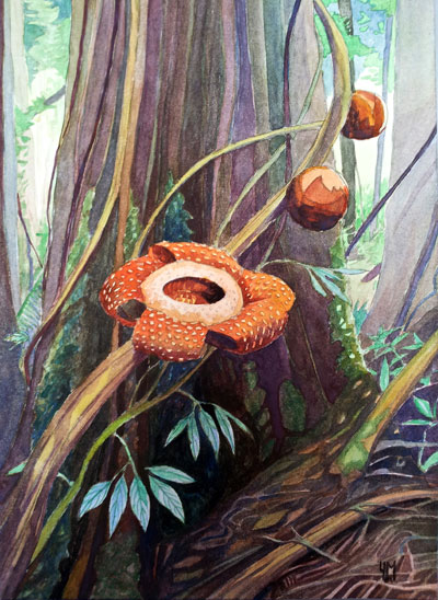
Rafflesia cantleyi (red flower) attached to it's host 
Tetrastigma rafflesiae. [9]

Within a DNA sequence there are transposable elements, which move segments of nucleic information elsewhere.These "jumping genes" can be a type of transposons called class 2 transposons. Type 2 transposons use a “cut and paste” function  so that they can “cut” a transposon out from their genome and then “paste” themselves into a target area, which could be either in a different segment of their own genome or a different genome altogether.2 An example of this can be seen between the parasitic plant Rafflesia cantleyi and it's host Tetrastigma rafflesiae. Researches have found that R. cantleyi has been retrieving genetic information from it's host to either gain nutrients more efficiently or to bypass the host's immune response to it.3 By taking advantage of transposons and integrating the two different genomes, parasitic R. cantleyi is able to improve its fitness and gives itself an edge over it's host.


RETROTRANSPOSONS

Transposable elements can also act as class 1 retrotransposons and use RNA as an intermediate between the two different genomes.4 An important parasite-host relationship that exploits the use of retrotransposons is the the HIV-human relationship. Human immunodeficiency virus is a retrovirus that uses RNA, reverse transcriptase, and integrase to fuse it’s own genetic information with the DNA of human cells. Once recombination occurs, the host's behavior is changed since it's own immune system begins to attack the infected cells, which are actually the body's immune cells. As the body attacks this hybrid of "host-parasite" cell, it also creates more immune cells, which HIV is patiently waiting to infect. This interaction allows HIV, the parasite, to continually infect other cells and create more copies of itself. This, in turn, increases HIV’s “fitness” due to an increase in reproductive success.


BACTERIOPHAGES

Parasites do not only exchange genetic information with their host, but also other symbiotic parasites that live in the same host. An example of this can be seen between different types of bacteria who take advantage of bacteriophages to integrate unique genetic information with one another.5 Bacteriophages help exchange DNA from bacteria such as, Staphylococcus aureus, to other cells to either continually reproduce the genetic information (lytic cycle) or to propagate its genetic information within its host species (lysogenic cycle).6 This recombination in the new parasitic cells can ultimately lead to resistance and increased efficiency in reproduction for the parasite.

The lytic and lysogenic cycle of bacteriophages which transfers genetic information from cell to cell. [10]


Why does it happen?

In many parasite-host relationship, an adaptive strategy made by either organism is done to improve fitness or to become a more efficient organism. When a parasite either gains or gives genetic information with it’s host, it is contributing in this exchange so that it has a more favorable environment and conditions to reproduce. The gene a parasite gives to its host could be one of antibiotic resistance, so that if the host tries to treat itself, it will prove ineffective. Likewise, a parasite could gain genetic information that prevents the host immune system from attacking it.7 Exchange of genetic information becomes an evolutionary tactic for the parasite to survive and to produce future generations. The parasite takes advantage of horizontal gene transfer in order to improve vertical gene transfer, and ultimately gives the parasite a more favorable opportunity to thrust its genes into the next generation.


SOURCES
  1. http://mbe.oxfordjournals.org/content/17/11/1769.full
  2. http://users.rcn.com/jkimball.ma.ultranet/BiologyPages/T/Transposons.html
  3. http://www.sciencedaily.com/releases/2012/06/120608100846.htm
  4. http://www.ebi.ac.uk/interpro/potm/2006_12/Page2.htm
  5. http://www.yale.edu/turner/pdf/p019.pdf
  6. http://onlinelibrary.wiley.com/doi/10.1111/j.1365-2958.2006.05354.x/pdf
  7. http://io9.com/5917214/parasitic-plants-actually-steal-the-genes-of-their-hosts
  8. http://users.rcn/com/jkimball.ma.ultranet/BiologyPages/T/Transposons.html 
  9. http://www.espacegraphique.com/blog/wp-content/uploads/2013/01/aquarelle-rafflesia.jpg
  10. http://classes.midlandstech.edu/carterp/Courses/bio225/chap13/img012.jpg
  11. http://www.youtube.com/watch?v=odRyv7V8LAE


Saturday, April 28, 2012

Mutualism: Women and L. acidophilus

Introduction: 


As human beings, we have thousands of different species of bacteria on our bodies, not including the other types of microorganisms. With all the different bacterial species, it would seem that no single bacteria is an indispensable member of our microbiome, however this is not the case. Lactobacillus acidophilus is a species of bacteria that women rely on just as much as the bacteria do. The L. acidophilus reside (among other places in the body) on the human vagina and use the woman as a resource host in exchange for protection from other potentially more harmful micro-organisms.

Description of the Relationship:


The presence of bacteria on the vagina was discovered over 100 years ago and is now one of the most well known examples of mutualism between humans and bacteria that there is. It is considered mutualistic because both the woman and the bacteria necessarily rely on one another. The L. acidophilus, once introduced to the vagina,  begins to adhere to the vaginal epithelial cells and colonize. The bacterial colony consumes the natural sugars excreted by the skin and ferments them into lactic acid. This product lowers the pH to a level intolerable by most bacteria not of the Lactobacillus genus. During menstruation, many of the bacteria are killed, reducing them to an appropriate level.


Cost/Benefit Analysis:


The woman benefits from this mutualistic relationship by gaining protection from more harmful organisms that could potential cause a yeast, bladder, or urinary tract infection. This is at the cost of a slight decrease in reproductive fitness. L. acidophilus taking glucose molecules that the woman worked to obtain might not have a large negative affect on the woman by itself, but there are thousands of micro-organisms on and inside the human body that all contribute to a somewhat heavy, yet necessary burden. The bacteria (L. acidophilus) benefit from the relationship by gaining a steady food supply, and a habitable living place. This is at the cost a attack by the woman's defense system (daily hygiene, monthly menstruation, and immune response if levels get too high)

References:


  1. "Lactobacillus acidophilus" University of Maryland Medical Center. http://www.umm.edu/altmed/articles/lactobacillus-acidophilus-000310.htm
  2. "Methods for Quantitative and Qualitative Evaluation of Vaginal Microflora During Menstruation" Pub Med Central. http://www.ncbi.nlm.nih.gov/pmc/articles/PMC238869/?tool=pmcentrez
  3. "Bacteria Genomes - LACTOBACILLUS ACIDOPHILUS" EMBL-EBI. http://www.ebi.ac.uk/2can/genomes/bacteria/Lactobacillus_acidophilus.html
  4. "Bugs Inside: What Happens When the Microbes That Keep Us Healthy Dissapear?" Scientific American. http://www.scientificamerican.com/article.cfm?id=human-microbiome-change
  5. http://www.youtube.com/watch?v=zJC5738poZE

Friday, April 27, 2012

Orchids and Orchid mycorrhiza


[6]
Introduction
            
            Orchids are widely distributed across the world with 25,000 to 35,000 species except for Antarctica [1].  Orchid’s lifecyle begins after pollination with the shivering of its petal and sepals. Then, it begins to form a seed; The maturation period of the seed is unique to each orchid species.  Next, the seed germinates into a seedling resulting in the flowering of orchid and the process begins again.  Since orchids’ seeds lack endosperm, they use resources from fungus to complete the germination process [2]. 


Description of the Relationship
Intracellular hyphal peloton [7]
            Basidiomycetes fungi provide energy and nutrients to orchids, from family Orchidaceae.  The fungi in the orchid mycorrhiza fulfill the role of endosperm in orchid seed [3].   During the non-photosythentic phase of the orchid’s lifecycle, the fungi provide the orchid with energy and carbon compounds [4].  After the non-photosynthetic phase, the fungi can also increase the nutrients and mineral intake of the orchid through the roots.   The fungi form pelotons that are like the host cell. These pelotons are surrounded by a membrane similar to plasma membrane. An infect orchid has altered microtubules and cell wall microfibrils [3].  All these alterations caused by the fungi help the orchid to absorb inorganic nutrients and trace elements from the soil.  The fungi also benefits from these interactions by receiving some of the photosynthesized carbohydrates [5]. Thus, the relationship between orchid and orchid mycorrhiza is mutualistic.  Some orchid mycorrhiza have evolved to be specific based on the species of the orchid therefore, can serve to be pathogenic to other orchid species [3].


Cost and Benefit Analysis
            As stated above, both the orchid and fungi benefit from this relationship.  The fungi gains carbohydrates from the orchid while the orchid receives nutrients and energy.  


The orchid does not spend a lot of energy in developing its roots in order to increase its volume of nutrients and minerals absorption from the soil.  Instead, the fungi fulfill that role.  The fungi are not limited to the roots alone.  They can explore other cavities and compounds that are not near the roots[5]. 
            Even though fungal infection can be pathogenic to the orchid, the orchid is able to control the infection.  The orchid has defense genes and compounds such as orchinol that can control the fungal infection [3].  These defense mechanisms does not significantly affect the reproductive success of the fungus.


References


Wednesday, April 25, 2012

Mighty Mitochondria and the Endosymbiosis Theory


Introduction

         Scientists approximate that bacteria first appeared on Earth around 3.5 billion years ago, while the first eukaryotes were not thought to inhabit Earth until approximately 1.5 billion years ago. What happened in this time gap of 2 billion years? Well, scientists can only make an educational guess, considering that primates likely did not come to be until approximately 65 million years ago (1). The endosymbiosis theory points to perhaps the earliest known form of mutualism in which bacteria are thought to have inhabited a “primordial eukaryotic cell” (2). The presence of a bacterial organism inside the larger cell allowed cellular life to adapt to the harsh conditions of the time. Scientists believe that photosynthetic bacteria appeared 3.2 million years ago, thereby releasing oxygen into the atmosphere. Oxygen can actually be a toxic substance, but the first aerobic bacteria lived about 2.5 billion years ago, very shortly before the first aerobic eukaryotes. These inferences, among many other observations, point to the endosymbiosis theory as a plausible explanation for adaption to an environment abundant in oxygen (1).

                                                                      Endosymbiosis Theory: http://www.youtube.com/watch?v=6DzzR76jj1k&feature=related 


Cost/Benefit Analysis          

          Why would we think that bacteria would crawl inside of another cell? Well, we assume that in a mutualistic relationship, both parties benefit due to the relationship. Additionally, the endosymbiosis theory suggests that mitochondria and chloroplasts’ lineages can be traced back to ancient bacteria. Not only are these structures about the size of a bacterium (70S), but these organelles also contain their own DNA in the form of one circular chromosome like bacteria (3). Both bacteria and the organelles replicate via binary fission (1).
Rickettsia prowazekki http://www.human-healths.com/rickettsia-prowazekii/rickettsia-prowazekii.php

         Although this background information is necessary in understanding the basic evolution of the eukaryotic cell, I would like to specifically examine the relationship between mitochondria (derived from ancient bacteria) and the primitive eukaryotic cell. As previously stated, at the time of the initial endosymbiotic encounter, few species were equipped to convert oxygen into another substance to avoid oxidation. Therefore, the host cell greatly benefitted from its smaller counterpart because mitochondria convert oxygen to ATP via substrate-level phosphorylation. Today, researchers have evidence to suggest the mitochondria are related to the proteobacterium Rickettsia prowazekii, “an obligate intracellular parasite” (3) due to its similarity in genomic sequence. Although it is not exactly clear why the bacterium benefitted from the relationship, it can be inferred that the bacterium was provided a “safe” environment to reside in a chemically stable environment. This idea applies to chloroplasts as well.

Mitochondria and the Cell

http://micro.magnet.fsu.edu/cells/mitochondria/mitochondria.html
         To understand the role of early bacteria in the cell, let us look at the function of a mitochondrion. Ultimately converting oxygen to ATP, mitochondria are the powerhouses of a cell through their production of energy. Mitochondria have two membranes, of which the outer membrane, containing porins, resembles Gram-negative bacteria. The inner membrane contains cristae, infoldings that greatly increase surface area. The inside of the mitochondria, the mitochondrial matrix, contains ribosomes (similar to size of those of bacteria), DNA, and calcium phosphate granules. The DNA and ribosomes are used to synthesize some of its proteins (3).

        The mitochondrion is the location of the tricarboxylic acid cycle (TCA), production of ATP, and oxidative phosphorylation.  In the TCA cycle, pyruvate is oxidized and cleaved to form CO2 and acetyl-coenzyme A (acetyl-CoA), an energy-rich molecule. After a series of reactions on acetyl-CoA, NADH and FADH2 are formed, which are both electron carriers. This reaction also yields four ATP molecules. The electron carriers formed in the TCA cycle are active players in the electron transport chain, as electrons are transferred to acceptors such as oxygen. The electron transport chain involves an energy gradient within the inner membrane of the mitochondria in eukaryotes and in the plasma membrane in prokaryotes. In this process, NADH is oxidized to NAD+, while the electrons are transferred to other molecules (such as oxgygen) with more positive reduction potentials. Oxidative phosphorylation is the result of the electron transport chain, in which protons move across the mitochondrial membrane and ultimately produce a maximum of 34 ATPs. These processes are very simplified, as more complex explanations involve in-depth biochemistry (3).

         Through these complicated processes in the mitochondria, eukaryotic cells are able to reap the benefits of an abundant amount of energy to sustain more complex forms of life. Although the TCA cycle and electron transport chain produce small amounts of ATP, oxidative phosphorylation allows for 34 molecules of ATP to be produced.

Mitochondria and the Body

An eye that has been affected by optic neuropathy.
 http://www2.cfpc.ca/cfp/2003/Oct/vol49-oct-clinical-2.asp
          We already have an idea of how beneficial mitochondria are to sustaining life, but what happens when mitochondria start to malfunction? As already mentioned, mitochondria contain their own DNA. Mitochondria are passed from mother to offspring in the cytoplasm of the egg. Over 200 human diseases are caused by mutations in DNA due to mitochondrial DNA’s inability to repair, unlike nuclear DNA. Most of these diseases come about in cells that require an abundance of ATP, such as muscle and nerve cells. Several examples of dysfunctional mitochondria-caused diseases are the following: optic neuropathy, often resulting in blindness; neurogenic muscle weakness; maternal and cardiomyopathy; and myoclonic epilepsy (2). Regardless of the primitive bacteria-eukaryotic cell relationship, it is apparent that eukaryotic cells have become very dependent on mitochondria to sustain life. 


References

    (1) The Endosymbiotic Theory. (2004, February 18). Retrieved from http://www.biology.iupui.edu/biocourses/N100/2k4endosymb.html.
    (2)    Brooker, R. J. (2009). Genetics: Analysis & Principles. (4 ed., pp. 119-120). New York: McGraw Hill. Print.
    (3)  Willey, Sherwood, and Woolverton. (2008).  Microbiology.  (7 ed., pp. 88 and 476-478). New York: McGraw Hill. Print.

Sunday, April 22, 2012

Farming beetles: Ambrosia beetles and their fungus

Different ambrosia beetles from tropical rainforests.
Introduction:
As we have seen with the Attine ant and Leucoprini fungus, animals have beaten humans to the act of farming. Another example of this would be the ambrosia beetles and ambrosia fungi. Ambrosia beetle is a general term used to describe several species of beetles from the two weevil subfamilies Scolytinae and Platypodinae [1]. One of the most rapidly diversifying, most widely distributed, and probably most ecologically and economically important group of ambrosia beetles is the tribe Xyleborini [1]. Ambrosia beetles can be found in all regions with tropical vegetation and even in some temperate areas [1].


Beautiful pink Fusarium repeatedly isolated from mycangia of Xylosandrus crassiusculus.


Since there are several species of ambrosia beetles, it comes with no surprise that there are many species of fungi that have a long established relationship with these beetles. The most common are polymorphic asexual anamorphs from the general Ambrosiella, Raffaelea, Ambrosiozyma and Dryadomyces [1]. Most ambrosia fungi are not capable of living independently of the beetles [1].


Description of the relationship:

http://www.ambrosiasymbiosis.org/
The relationship between the beetles and the fungus has been describes as one of the most successful and widespread cases of insect-fungus symbiosis, ambrosia beetles are the product of sixty million years of intricate mutualism between the faster evolving beetles and some of the most bizarre fungi [2].

Surface pit mycangia. Left: Female of Treptolatypus solidus with glandular pit mycangia on top of the pronotum. Right: Simple nonglandular pit mycangium on the head of Scolytodes unipunctatus. Arrow on the right points to a fungus spore.
Ambrosia beetles have evolved a variety of morphological structures to carry their fungal symbionts from tree to tree. These structures are generally called mycangia [1]. As the beetles leave their initial home, they carry spores of the fungus in their mycangia. Once they find a suitable tree, they then create tunnels (galleries)  inside the wood which will make small strings of compacted sawdust protrude from the tree [3].

Sawdust stacks on a dead tree - typical sign of ambrosia beetles at work.
The beetles will then cultivate the fungi inside the galleries within dead or decaying wood and feed on their mycelium. Once the "fungus farming" has started, the beetles can lay their eggs that will produce larva. The beetles will care for their young larva in the trees where they will become beetles and carry fungal spores to their next habitat.

Cost/benefit analysis:
     The beetles are dependent upon the fungi, from which they require amino acids, vitamins, and sterols [5]. Certain immature stages of the beetles are unable to complete development in the absence of the fungus [6], At the same time, the activities of female beetles have been hypothesized to control the growth of composition of ambrosial gardens. If the female dies, the garden is quickly overgrown by contaminating fungi and bacteria, which ultimately results in the death of the brood [5].
     The interaction is clearly mutualistic. The symbiosis allows the beetles to exploit a nutritionally poor resource (wood) and reduce interspecific competition, while providing the fungi consistent transport to a relatively rare and ephemeral resource (a new host of the appropriate condition and successional stage) [5].
     Although both the beetle and the fungi are benefited by this relationship, there are also cost to pay. The beetle could die if the fungus is suddenly removed from its habitat as the beetles are too specialized to be able to use another species of fungus. The fungus has also evolved to where it does not reproduce sexually, thus reducing genetic diversity and ability to explore different niches without the help of the beetle.

References:
[1] http://www.ambrosiasymbiosis.org/
[2] http://itp.lucidcentral.org/id/wbb/xyleborini/
[3] http://edis.ifas.ufl.edu/hs379
[4] http://www.mapoflife.org/topics/topic_137_Beetles-insights-into-convergence/
[5] http://www.mdpi.com/2075-4450/3/1/339/
[6] http://ddr.nal.usda.gov/bitstream/10113/9868/1/IND22088830.pdf

Saturday, April 21, 2012


Living Easy: Podarcis lilfordi and Daphne rodriguezii


                                   http://www.arkive.org/lilfords-wall-lizard/podarcis-lilfordi/

Introduction:

The lizard, Podarcis lilfordi, is found on the Balearic Islands off the coast of Spain [1]. However, unlike other lizards, this species shares a mutualistic relationship with the shrub Daphne rodriquezii [2]. D. rodriquezii is characterized from its beautiful white flowers and it also contains many fruits that the lizards like to consume [3]. The lizards consume and disperse the seeds of the plant by eating the fruits that grow from the shrubs [4].

 P. lilfordi is a medium sized lizard that grows to a maximum of 80mm. Its body mass can be anywhere between 4.2 to 9.5 grams. The females reproduce cyclically, producing 2-4 eggs per clutch. They can lay multiple clutches in one season [3]. However, as new predators and lizards show up to the islands and compete for resources, the population of P. lilfordi has slowly started to decline [1]. The shrub, D. rodriguezii, starts to grow during the early spring season. They contain very small, white flowers. The sepals and petals unite, forming a tube called the hypanthium that usually has purple dyes and is 7 to 11 mm in length. During the summer, the shrubs mature and produce red-orange, fleshy globose fruits. The shrubs rarely grows above 50 centimeters in height [2].


         http://mundani-garden.blogspot.com/2012/04/daphne-rodriguezii-art-of-camouflage.html

Description of the Relationship:
P. lilfordi are very productive seed-dispersers for the shrubs. It is a mutualistic relationship, in which the lizards use the shrubs not only as a food source, but also as a second home [1]. The lizard eats the fruits that D. rodriguezii produces, and it also uses the shrub as a home, keeping it out of sight from other predators and the hot sun [2]. In return, the shrub gets the benefit of having the lizards disperse their seeds for them. They are then better able to spread their offspring to other areas [3].
However, P. lilfordi also has other priorities and responsibilities on the ecosystem found on the Balearic Islands. It is also a pollinator of the aroid Dracunculus muscivorus, and a seed disperser for Withania frutescens and Phillyrea media [1].

Cost Benefit Analysis:


As mentioned before, the mutualistic association between P. lilfordi and D. rodriguezii is very straightforward. D. rodriguezii serves as both a food source and a shelter for P. lilfordi, and in return P. lilfordi disperses the seeds of the shrub through fecal defecation. Without D. rodriguezii being present in nature, P. lilfordi would still be able to survive and reproduce in nature. However, it would have a better chance to pass its progeny if D. rodriguezii was present, as it would have an extra food source and shelter [1]. D. rodriguezii, however, would have a much harder time reproducing in nature as their seeds would not get dispersed if P. lilfordi was not present in the environment.  


References:


Relaxing in the Rectum


Introduction:

  http://animals.nationalgeographic.com/animals/invertebrates/sea-cucumber/
Many organisms living in the sea benefit from a symbiotic relationship with other organisms around them. One example of this is the relationship between the sea cucumber and the pearl fish. Sea cucumbers are echinoderms that have a shape very similar to soft cucumbers (1). They have “leathery skin” and their body is essentially one long digestive tube. These invertebrates crawl along the floor of the ocean and can reside in both shallow and deep areas of the ocean. They are omnivorous and usually eat small particles and detritus they find on the ocean floor. The sea cucumber can grow between 2 to 200 cm and can live to be between 5 and ten years old (2). Spawning for the sea cucumber usually occurs between June and August. The sea cucumbers release their sperm and eggs in the water and allow fertilization to take place. These fertilized eggs continue to develop into larvae, which become suspended in the water column for up to 70 days. Eventually, these planktonic forms settle on the ocean floor, where they change into tiny little juvenile sea cucumbers. Growth is slow for these invertebrates, and it can take up to 4 years for the cucumber to reach adult size (3).  
http://www.bing.com/images/search?q=pearl+fish&view=detail&id=15EA9DE49EDFD35990AFEEFBD2FA3E98694E81B7&first=0&qpvt=pearl+fish&FORM=IDFRIR

The symbiont of the sea cucumber is the pearl fish. Pearl fish are “eel-shaped” fish that are found worldwide, but primarily in tropical, shallow areas around coral reefs. They have long, slender bodies that lack scales and they usually have transparent skin. The tail of the pearl fish is long and pointed, and the anus of this fish is located close to its neck. Pearl fish typically grow to be about 15 cm long (4). The female pearl fish releases clumps of eggs late in the summer to begin the life cycle. These eggs rise to the surface and hatch, turning into a specific type of larvae called vexillifers. These larvae live among the plankton until reaching a length of about 7 to 8 cm. At this point, the larvae develop into tenuis. These forms descend to the ocean floor and begin their search for food and a host (5). The pearl fish will constantly be on the lookout for sea cucumbers to create a symbiotic relationship.


http://www.youtube.com/watch?v=jM9Y4ww2O_s&feature=related

Description of Relationship:
Once a pearl fish (Onuxodon or Carapus) finds a sea cucumber (Holothuroidea), it immediately begins to smell around to distinguish between the head and the anus of the cucumber (6). Once it finds the anus, the pearl fish works its way into the rectum of the sea cucumber, eventually being completely engulfed in the digestive canal of its host. There it will spend the day inside, using its host as a form of protection. At night, the pearl fish comes out to feed on small crustaceans, but it doesn’t go too far from its host (7). After feeding, the pearl fish returns to its host and waits for the sea cucumber to take a breath. When the anus opens for respiration, the pearl fish simply swims back inside, seeking shelter in the rectum of its host (8). The pearl fish and the sea cucumber have evolved a symbiotic relationship know as commensalism. In this relationship, the pearl fish benefits because it gains a place to live that is cozy and protected from predators as well as any nutrients that can be absorbed as they flow out of the cucumber’s anus. Meanwhile, the sea cucumber appears to be unaffected by this relationship. It doesn’t even seem to notice the pearl fish entering its anus. As far as we know, the pearl fish is not taking anything from the sea cucumber. The reproductive success of the cucumber remains the same. Therefore, since the pearl fish benefits and the sea cucumber is neither helped nor harmed, one can argue that this relationship is one of commensalism. Many other organisms have benefitted from relationships similar to that of the sea cucumber and the pearl fish. The pearl fish have also learned to penetrate the bodies of other invertebrates such a starfish, sea squirts, and clams. A number of crabs and polychaete worms have also evolved to live inside sea cucumbers and have become specialized for gaining protection from the cloaca of that host (9).

http://www.merchantcircle.com/blogs/Kinetic.Illusions.Creative.Studio.And.Video.Production.916-706-1058/2010/7/Rare-footage-of-a-pearlfish-penetrating-the-anus-of-a-sea-cucumber/588295

Cost/Benefit Analysis:
The relationship between the pearl fish and the sea cucumber is not obligatory, but the pearl fish benefits from its symbiosis with the sea cucumber. The costs for the pearl fish in this relationship are very small. They must expend energy searching for a sea cucumber and for wiggling their way into the anus of the host. On the other hand, the benefits for the pearl fish are immense. The fish is provided protection from predators when it resides within the rectum of the cucumber. They also can take up any nutrients found in the waste products being excreted from the rectum of the sea cucumber. Another benefit for certain species of pearl fish is that they can use the rectum of the sea cucumber as a place to develop into their adult forms and complete their life cycle (9). While the pearl fish is provided with shelter, the sea cucumber has nothing to gain from this relationship. Nor does it have anything to lose. Both the cost and benefit for the sea cucumber is nothing. The reproductive success of the sea cucumber is not affected and the pearl fish benefits, it is likely that this relationship will persist as commensalism. Eventually, there may even be an adaptation that would benefit the sea cucumber, creating a relationship that is more like mutualism.


References:
1. http://blogs.thatpetplace.com/thatfishblog/2008/11/26/pearlfish-and-sea-cucumber-symbiosis/
2. http://animals.nationalgeographic.com/animals/invertebrates/sea-cucumber/
3. http://bioweb.uwlax.edu/bio203/s2008/hui_ka/04Life_Cycle.html
4. http://www.britannica.com/EBchecked/topic/448066/pearlfish
5. http://www.britannica.com/EBchecked/topic/442415/paracanthopterygian/63545/Natural-history?anchor=ref525827
6. http://www.fishbase.org/Summary/FamilySummary.cfm?Family=Carapidae
7. http://www.ms-starship.com/sciencenew/symbiosis.htm
8. http://www.reefed.edu.au/home/explorer/animals/marine_invertebrates/ echinoderms/sea_cucumbers
9. http://en.wikipedia.org/wiki/Sea_cucumber